Varicocele Grades 1, 2 & 3: Symptoms, Doppler & Treatment
Varicocele Grading Guide

Varicocele Grades 1–5: Symptoms, Doppler Grading & Treatment Guide

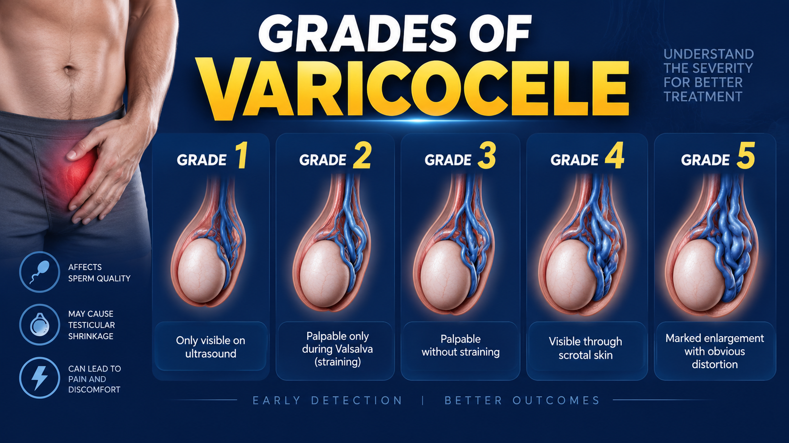
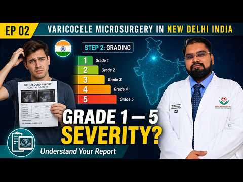
Varicocele grading becomes confusing because two systems are commonly discussed. On physical examination, doctors use clinical Grade 1, Grade 2 and Grade 3. On ultrasound, Doppler systems such as Sarteschi Grade 1–5 describe where reflux is seen and how the veins behave.

Do not convert millimetres directly into a clinical grade. A 3 mm or 4 mm vein can support the Doppler diagnosis when reflux is present, but clinical Grade 1, 2 or 3 is decided by examination.
Dr. Vijayant Govinda Gupta
Clinical Varicocele Grading

Clinical Varicocele Grades 1, 2 and 3

Clinical varicocele grading is based on what can be felt or seen while the patient is standing. This is separate from Doppler vein size and separate from Sarteschi ultrasound Grade 1–5.

Varicocele symptoms, testicular size and warning signs explained
1

Varicocele Grade 1

Examination: felt only during the Valsalva manoeuvre. Symptoms: often none; some men notice mild heaviness or a dull ache after prolonged standing or exercise. Fertility: grade alone does not predict sperm quality. Treatment: usually considered only when there is a relevant problem such as abnormal semen parameters with infertility, persistent typical pain, or objective testicular impact.

2

Varicocele Grade 2

Examination: palpable while standing at rest, but not clearly visible through the scrotal skin. Symptoms: heaviness or dragging discomfort may be more noticeable, although some men remain symptom-free. Fertility: semen analysis is more useful than grade alone. Treatment: observation is reasonable when symptoms and fertility findings are reassuring; repair may be discussed when pain, fertility or testicular size is affected.

3

Varicocele Grade 3

Examination: visible and palpable at rest, often described as a “bag of worms”. Symptoms: pain and heaviness can occur but are not universal. Fertility: a proper semen analysis and testicular-volume assessment are useful when fertility matters. Treatment: a large grade alone is not an automatic reason for surgery; the decision depends on symptoms and objective testicular or fertility findings.

What about a subclinical varicocele? A varicocele that is seen only on ultrasound and cannot be felt on examination is usually described as subclinical. It should not be confused with clinical Grade 1. For male infertility, major guidelines generally do not recommend repair of an imaging-only non-palpable varicocele.
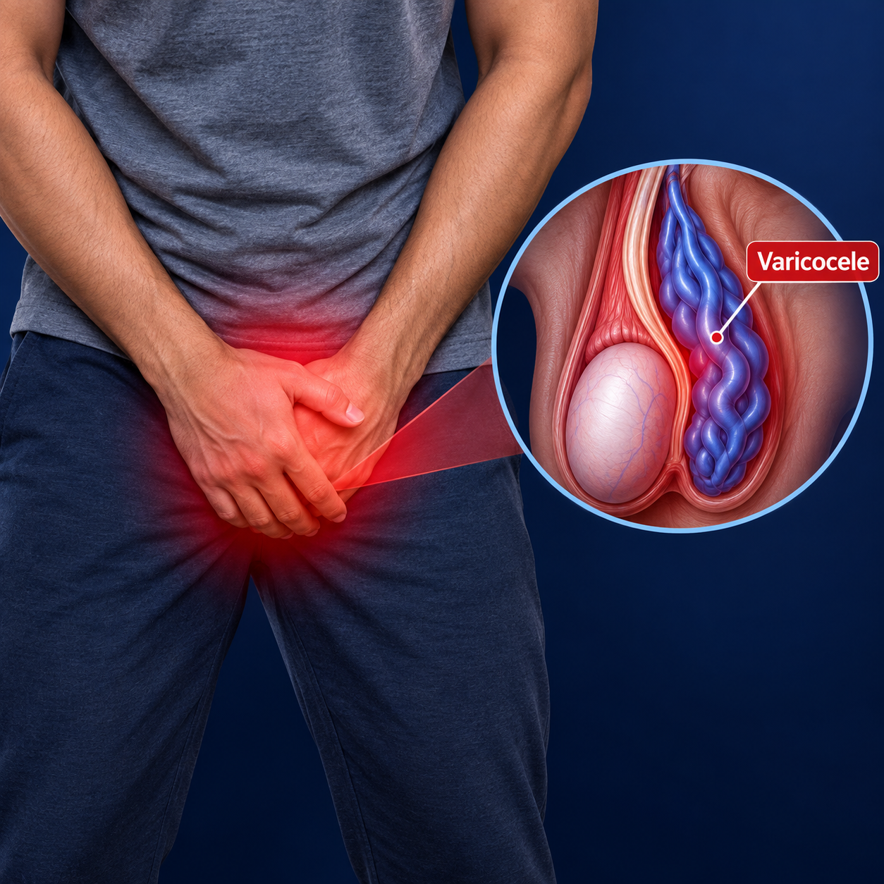
Common varicocele symptoms and warning signs
Varicocele Symptoms

Common Symptoms and Findings Patients Notice

The grade and the symptoms do not always move together. A small varicocele can ache, while a large one may cause little discomfort.

Dull dragging ache

Usually worse after long standing, exercise or late in the day, and often improves when lying down.

Scrotal heaviness

A feeling of weight or pulling around the affected testicle, especially during activity.

Visible enlarged veins

More obvious in larger clinical varicoceles and commonly described as a “bag of worms”.

Difference in testicular size

More relevant in adolescents and younger men, where serial testicular-volume measurements may be useful.

Abnormal semen analysis

Some men with a clinical varicocele have reduced sperm concentration, motility or other semen abnormalities.

No symptoms at all

Many varicoceles are found during fertility assessment or a routine examination rather than because of pain.

Varicocele Doppler Grading

Sarteschi Doppler Grades 1–5

Sarteschi grading describes the ultrasound pattern of venous reflux, the position of the abnormal veins and whether testicular hypotrophy is present. It is an ultrasound classification and should not be used as a replacement for clinical Grade 1, 2 or 3.

Varicocele Doppler ultrasound grading, reflux and vein diameter
Sarteschi gradeTypical Doppler patternWhere reflux is seenTesticular finding
Grade IReflux appears during Valsalva; obvious scrotal varices are not seen on routine imaging.Mainly inguinal canal.No testicular hypotrophy expected from the grading definition.
Grade IISmall supratesticular varicosities with reflux during Valsalva.Above the upper pole of the testis.Usually no hypotrophy in the grading definition.
Grade IIIVaricosities extend around/towards the lower pole and reflux is provoked by Valsalva, especially standing.Peritesticular / lower scrotal veins.Hypotrophy is not a defining feature.
Grade IVDilated veins can be seen even supine. Reflux is present at rest and increases with standing or Valsalva.Peritesticular veins.Reduced testicular volume may be present.
Grade VDilated veins are present in multiple positions, with spontaneous resting reflux that shows little or no further increase with Valsalva.Extensive pampiniform plexus reflux.Testicular hypotrophy is commonly associated in the classic description.
Why the distinction matters: “Clinical Grade 2” and “Sarteschi Grade 2” describe different things. One is based on what the doctor feels; the other is an ultrasound pattern.
Varicocele Vein Size in mm

What Does a 2.5 mm, 3 mm, 4 mm or 5 mm Varicocele Mean?

This is one of the most common points of confusion on Doppler reports. Diameter helps, but reflux and examination matter more than a single number.

2.5 mm

Borderline measurement

A 2.5 mm vein can be seen in some normal or borderline studies. By itself, it does not prove clinically significant varicocele.

3 mm

Common ultrasound threshold

A maximum vein diameter above 3 mm, especially measured standing during Valsalva, supports the diagnosis when significant reflux is also demonstrated.

4 mm

Clearly dilated vein

A 4 mm varicocele vein is enlarged, but “4 mm” still does not tell you whether the clinical grade is 1, 2 or 3.

5 mm

Larger venous dilation

A 5 mm vein is markedly dilated. Management still depends on reflux, symptoms, testicular size, semen findings and physical examination.

Evidence-based interpretation: EAU and ESUR guidance supports measuring the largest vein while standing and during Valsalva. A maximum diameter above 3 mm together with reflux lasting more than 2 seconds is a useful pattern for clinically significant varicocele. Different studies have used different diameter cut-offs, so a Doppler report should never be interpreted from millimetres alone.
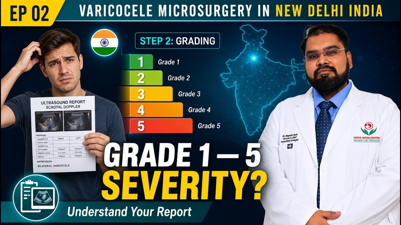
How to Read a Varicocele Doppler Report

How to Read a Varicocele Doppler Report

A useful report should describe the side involved, vein diameter, reflux, patient position and the volume of both testes. These details are more useful than a vague label such as “mild” or “moderate”.

1. Side: left, right or bilateral

Most varicoceles are left-sided, but both sides should be examined and reported when relevant.

2. Largest vein diameter

Ideally measured in the upright position during Valsalva. The exact technique matters because the diameter can change with posture and straining.

3. Reflux duration

Reflux means reverse venous flow. ESUR suggests that reflux lasting more than 2 seconds while standing during Valsalva is abnormal.

4. Reflux at rest vs only with Valsalva

Spontaneous resting reflux generally represents a more haemodynamically advanced Doppler pattern than reflux that appears only with straining.

5. Testicular volume on both sides

Volume should be compared side by side. EAU guidance recommends the Lambert formula: length × width × height × 0.71.

6. Location of the dilated veins

Inguinal, supratesticular and peritesticular location helps when a Sarteschi Doppler grade is being assigned.

Example of a useful Doppler statement

Left pampiniform plexus veins measure up to 3.6 mm while standing during Valsalva. Retrograde flow persists for 2.8 seconds. Left testicular volume 15 mL; right testicular volume 16 mL.

This tells us the vein is dilated and there is significant reflux, while testicular volumes are fairly similar. The clinical Grade 1, 2 or 3 still comes from physical examination.

Real Patient Questions

Varicocele Grades FAQs

Clear answers to common questions about grades, Doppler reports, pain, fertility and treatment — with the most relevant doctor-video chapter beside each answer.

Yes. Varicocele can still show on ultrasound after surgery, and varicocele can also recur. Enlarged veins may remain visible even when the abnormal reflux has stopped, so recurrence is judged by symptoms, physical examination and whether meaningful reflux persists on Doppler. A vein that is still visible on ultrasound does not by itself prove that the surgery has failed.

Why Does Varicocele Recur After Surgery? video
Relevant video answer
Why Does Varicocele Recur After Surgery?
Starts at 00:00

No. Every varicocele grade does not need surgery. Clinical Grade 1, Grade 2 or Grade 3 is only one part of the treatment decision. Surgery is usually discussed when a clinical varicocele is accompanied by infertility with abnormal semen parameters, persistent typical pain, or objective testicular impact. An imaging-only subclinical varicocele is generally not treated for infertility.

Do All Varicoceles Need Surgery? video
Relevant video answer
Do All Varicoceles Need Surgery?
Starts at 00:00

Yes. Varicocele can be associated with testicular shrinkage or reduced testicular volume, especially in adolescents and younger men. After successful treatment, some younger patients may show catch-up growth, but testis size improvement is not guaranteed. Serial examination and ultrasound measurements are more useful than judging recovery from a single scan.

Can an Atrophied Testicle Regain Size? video
Relevant video answer
Can an Atrophied Testicle Regain Size?
Starts at 03:30

If varicocele is present on both sides with different grades, each side should be assessed separately. The doctor considers the clinical grade, Doppler reflux, vein diameter, testicular volume, pain and fertility findings on the left and right sides. Treatment should not be based only on whichever side has the higher grade.

What Is Bilateral Varicocele? video
Relevant video answer
What Is Bilateral Varicocele?
Starts at 00:00

A higher varicocele grade can be associated with a greater chance of abnormal sperm count or motility in some men, but varicocele grade alone cannot predict male fertility. Semen analysis, testicular size, the female partner's fertility factors and the couple's timeline are more useful when deciding whether treatment may help.

Low Sperm Count & Infertility video
Relevant video answer
Low Sperm Count & Infertility
Starts at 05:35

To read a Doppler or ultrasound report for varicocele grading, check the side involved, maximum vein diameter, whether measurements were taken while standing and during Valsalva, reflux duration, reflux at rest, and the volume of both testes. A vein diameter above about 3 mm together with reflux lasting more than 2 seconds during standing Valsalva supports the diagnosis, but the report still needs clinical correlation.

Reflux Duration on Doppler (>2 Seconds) video
Relevant video answer
Reflux Duration on Doppler (>2 Seconds)
Starts at 04:50

Grade 1, Grade 2 or Grade 3 varicocele can all be associated with pain, and pain severity does not always match the grade. Typical varicocele pain is usually a dull dragging ache or heaviness that worsens with standing or activity. Other causes of scrotal pain should be excluded, and persistent typical pain despite conservative measures can be a reason to discuss repair.

Dull Dragging Pain in the Evening video
Relevant video answer
Dull Dragging Pain in the Evening
Starts at 01:40

Yes, varicocele can affect Army, NDA, Police or other medical fitness examinations, but the exact rules can change and may differ between services and recruitment cycles. Candidates should check the current official medical standards and keep their examination, Doppler and treatment records available for the medical board.

Army & Armed Forces Medical Standards for Varicocele video
Relevant video answer
Army & Armed Forces Medical Standards for Varicocele
Starts at 00:00

Yes. Grade 1 or Grade 2 varicocele can often be managed without surgery when symptoms are mild and semen parameters and testicular size are reassuring. Observation, scrotal support, avoiding activities that clearly trigger symptoms and appropriate pain relief may be enough. Conservative treatment can control symptoms, but it does not physically remove the dilated veins.

Grade 1 & Mild Varicocele Conservative Protocol video
Relevant video answer
Grade 1 & Mild Varicocele Conservative Protocol
Starts at 01:45

A 2.5 mm, 3 mm, 4 mm or 5 mm varicocele vein diameter does not directly convert into clinical Grade 1, Grade 2 or Grade 3. Clinical grading comes from physical examination. Vein diameter helps support the ultrasound diagnosis when reflux is present, but millimetres alone should not be used to assign a clinical grade.

Vein Diameter (mm) vs Clinical Symptoms video
Relevant video answer
Vein Diameter (mm) vs Clinical Symptoms
Starts at 04:30
Doppler Report Review

Not Sure What Your Varicocele Grade Means?

Discuss the clinical grade, vein diameter, reflux duration, testicular volume, semen report and symptoms together before deciding whether observation or treatment is appropriate.

+91-95929 99184
Scroll to Top